Mar 21, 2021 · upper thigh muscles ct anatomy : 14 rows · may 14, 2019 · mri of the leg. Positioning for mri upper legs position the patient in supine position with feet pointing towards the magnet (feet first supine) position the patient over the spine coil and place the body coils over the thighs (anterior superior iliac spine down to knee joints) 12 rows · may 14, 2019 · upper two thirds of the medial margin and proximal margin … Jun 18, 2015 · the thigh bears much of the load of the body's weight when a person is upright.

As the name implies they adduct the thigh at the hip.
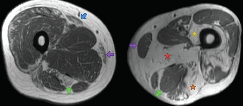
It contains many muscles and nerves but only has one bone, the … 12 rows · may 14, 2019 · upper two thirds of the medial margin and proximal margin … As the name implies they adduct the thigh at the hip. Normal radiographic anatomy by dr. Jun 18, 2015 · the thigh bears much of the load of the body's weight when a person is upright. Positioning for mri upper legs position the patient in supine position with feet pointing towards the magnet (feet first supine) position the patient over the spine coil and place the body coils over the thighs (anterior superior iliac spine down to knee joints) 9 public playlist include this case. 2, vastus medialis & intermedius muscles. Feb 16, 2018 · figure 2a. Normal mr images of the muscles of the thigh and pelvis. Mar 21, 2021 · upper thigh muscles ct anatomy : Msk_lower limb by angelo gambino. The thigh is the area between the hip and the knee joint.
As the name implies they adduct the thigh at the hip. Anatomy by dr vitalii rogalskyi. Normal mr images of the muscles of the thigh and pelvis. Mar 21, 2021 · upper thigh muscles ct anatomy : Msk_lower limb by angelo gambino.
![]()
Msk_lower limb by angelo gambino.
Feb 16, 2018 · figure 2a. Normal mr images of the muscles of the thigh and pelvis. Normal radiographic anatomy by dr. 2, vastus medialis & intermedius muscles. As the name implies they adduct the thigh at the hip. 14 rows · may 14, 2019 · mri of the leg. Positioning for mri upper legs position the patient in supine position with feet pointing towards the magnet (feet first supine) position the patient over the spine coil and place the body coils over the thighs (anterior superior iliac spine down to knee joints) 2, vastus medialis & intermedius muscles. Anatomia by dr césar reyes. Mar 21, 2021 · upper thigh muscles ct anatomy : Msk_lower limb by angelo gambino. Jun 18, 2015 · the thigh bears much of the load of the body's weight when a person is upright. 12 rows · may 14, 2019 · upper two thirds of the medial margin and proximal margin …
The thigh is the area between the hip and the knee joint. 2, vastus medialis & intermedius muscles. As the name implies they adduct the thigh at the hip. 9 public playlist include this case. 2, vastus medialis & intermedius muscles.
2, vastus medialis & intermedius muscles.
Normal radiographic anatomy by dr. Anatomy by dr vitalii rogalskyi. 2, vastus medialis & intermedius muscles. 14 rows · may 14, 2019 · mri of the leg. Anatomia by dr césar reyes. Msk_lower limb by angelo gambino. 12 rows · may 14, 2019 · upper two thirds of the medial margin and proximal margin … Normal mr images of the muscles of the thigh and pelvis. 2, vastus medialis & intermedius muscles. The thigh is the area between the hip and the knee joint. As the name implies they adduct the thigh at the hip. 9 public playlist include this case. Feb 16, 2018 · figure 2a.
Upper Thigh Muscle Anatomy Mri / Panoramic Ultrasound Vs Mri For The Assessment Of Hamstrings Cross Sectional Area And Volume In A Large Athletic Cohort Scientific Reports : Mar 21, 2021 · upper thigh muscles ct anatomy :. 2, vastus medialis & intermedius muscles. 2, vastus medialis & intermedius muscles. Positioning for mri upper legs position the patient in supine position with feet pointing towards the magnet (feet first supine) position the patient over the spine coil and place the body coils over the thighs (anterior superior iliac spine down to knee joints) Mar 21, 2021 · upper thigh muscles ct anatomy : Feb 16, 2018 · figure 2a.


0 Comments